Animal healthcare is becoming more preventive, mobile, and evidence-driven. The American Pet Products Association’s 2023–2024 National Pet Owners Survey reports that 66% of U.S. households own a pet. This expanding patient population increases demand for faster, practical diagnostic support.
A Portable Animal Ultrasound Machine brings imaging closer to the examination table, farm pen, or emergency vehicle. A clinician can scan a dog with abdominal pain without moving it across a busy hospital. A field veterinarian can examine a pregnant mare beside a stable. Small details matter. Battery operation, probe selection, image storage, and cleaning design influence daily performance.
Grand View Research identifies portable systems as an important direction within the veterinary ultrasound market. MarketsandMarkets also describes growing demand for veterinary diagnostic imaging, supported by rising pet care spending and expanding animal health services. These reports suggest strong momentum, but market growth does not guarantee clinical quality. That part needs careful thought.
Ultrasound remains operator-dependent. A lightweight device cannot replace anatomical knowledge, patient preparation, or professional interpretation. The American College of Veterinary Radiology emphasizes appropriate training and diagnostic review when imaging findings are uncertain. In practice, the best system is not always the one with the highest advertised specifications. It should produce clear images, withstand repeated handling, and fit the clinician’s workflow.
Portability can improve access and speed. It can also expose weak training. That is the uncomfortable point.
Choosing wisely means comparing image quality, ergonomics, software, service support, and total ownership costs. The right Portable Animal Ultrasound Machine supports informed decisions while keeping animal welfare at the center.
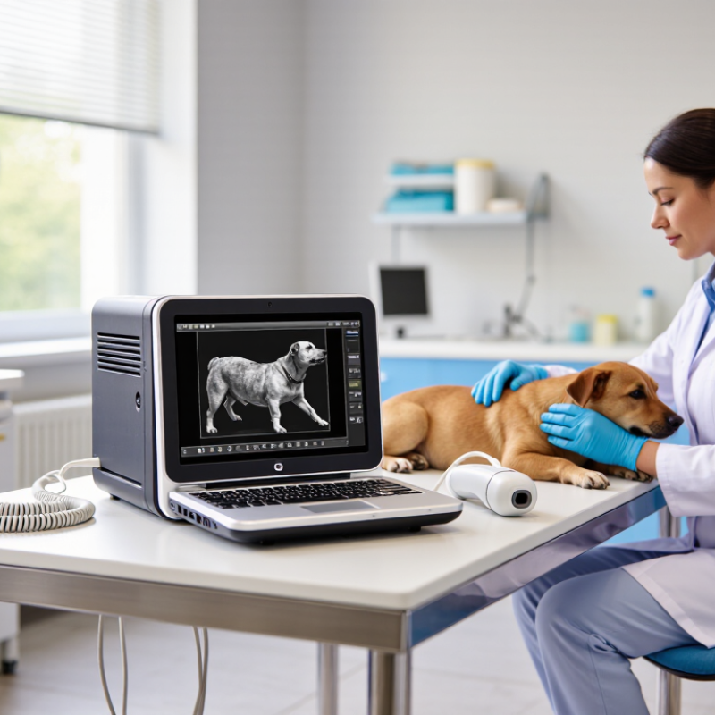
A portable animal ultrasound machine is a compact diagnostic system that creates real-time images using high-frequency sound waves. It does not use ionizing radiation. A veterinarian can examine a horse’s tendon, a dog’s abdomen, or a pregnant ewe beside the treatment table. The system usually includes a handheld probe, a display, rechargeable power, and image-storage software. Some models weigh only a few kilograms, so one person can carry the unit between rooms or farms.
This mobility matters in everyday veterinary work. A 2024 Grand View Research report estimated the global veterinary ultrasound market at approximately USD 190 million in 2023, with continued growth expected through the decade. MarketsandMarkets also identified point-of-care diagnosis as a major market driver in its 2023 analysis. These figures reflect rising demand for faster imaging, but they do not guarantee better clinical decisions. Training still matters.
The probe sends sound into tissue and receives returning echoes. Software converts those echoes into moving images. Different probes suit different tasks. A linear probe may show a superficial tendon, while a convex probe can reach deeper abdominal organs. In practice, clipped hair, nervous animals, thick fat, or poor positioning can reduce image quality. Portable does not mean effortless. An experienced operator still needs patient restraint, correct settings, and clinical judgment. I would question any scan interpreted without the animal’s history, physical examination, and follow-up evidence.
Portable animal ultrasound machines typically weigh far less than cart-based systems, making them easier to carry between examination rooms, farms, stables, and field locations. Lower equipment weight can improve mobility while supporting faster point-of-care examinations. Actual weight varies according to the console, battery, display, and transducer configuration.
Portable ultrasound supports veterinary diagnosis by bringing imaging closer to the animal. A veterinarian can examine a restless dog in the consultation room, rather than moving it across the clinic. This reduces handling stress and saves valuable time. A small probe can reveal bladder stones, abdominal fluid, intestinal changes, or pregnancy-related findings. In farm settings, it can also support reproductive checks beside a stall or pasture gate.
Clear images still depend on training, probe selection, and patient positioning. A clipped coat and a thin layer of gel improve contact. For example, a calm cat lying on a padded table may provide better images than a restrained cat in a noisy room. Ultrasound does not replace blood tests, radiographs, or clinical judgment. Some findings remain uncertain, and poor positioning can mislead even an experienced operator. That limitation deserves honest attention.
Tips: Keep the machine charged and disinfect the probe after every examination. Use species-appropriate presets when available. Record the animal’s position, scanned area, and key findings. Compare images with physical signs and medical history. When an image seems unclear, ask a qualified veterinary professional to review it or recommend further testing. A portable system is useful, but it is not automatically accurate.
Why Choose a Portable Animal Ultrasound Machine?
Field ultrasound brings real-time evidence to farms, stables, and remote clinics. A technician can examine pregnancy, ovaries, tendons, lungs, or abdominal organs beside the animal. No transport is needed. That reduces handling stress and saves valuable time.
FAO reports that livestock support the livelihoods of about 1.3 billion people worldwide. In these settings, earlier decisions can protect animal health and household income. The USDA APHIS NAHMS Dairy 2014 study also identifies reproductive monitoring as a major management activity on dairy operations. Ultrasound adds detail that touch alone may miss. It can show fluid, follicles, fetal movement, or tissue changes. The image is immediate. Still, it is not magic. Poor restraint, dirty probes, or limited training can produce misleading results. I would always compare the scan with history, physical findings, and follow-up testing.
Tips: Choose a unit with a sealed probe, readable screen, and dependable battery life. Practice on healthy animals before scanning difficult cases. Record the date, body region, findings, and operator. Keep images when possible. Review uncertain scans with a qualified veterinarian. The British Veterinary Association stresses that diagnostic tools should support, not replace, professional clinical judgment. One overlooked detail can matter. Field work remains imperfect.
Why Choose a Portable Animal Ultrasound Machine?
Choosing a portable animal ultrasound machine starts with the examination setting. In a barn, I often need one hand free while checking a restless mare. Weight, grip comfort, and screen visibility matter more than impressive specifications. A 2024 Grand View Research estimate valued the global veterinary ultrasound market at roughly USD 300 million in 2023, reflecting growing demand for accessible diagnostic imaging. Market growth alone should not dictate a purchase.
Probe selection deserves careful attention. A microconvex probe can support abdominal examinations, while a linear probe may show tendons and superficial structures more clearly. Confirm the available frequency range, depth, frame rate, and image storage format. The device should also offer reliable battery performance, dust resistance, and simple cleaning between patients. Battery claims can be optimistic. Real use depends on brightness, scanning mode, and wireless functions.
Independent testing is valuable. The FDA explains that diagnostic ultrasound does not use ionizing radiation, but appropriate operator training remains essential. A 2023 review in Veterinary Radiology & Ultrasound emphasized that image quality and clinical interpretation depend strongly on user experience. Ask about service response times, software updates, probe replacement costs, and local technical support. DICOM compatibility may help hospitals maintain organized records. A lower purchase price can become expensive when repairs, accessories, and training are overlooked. My own weak point is convenience bias; a lighter unit is not automatically the better clinical tool.
| Factor | Why It Matters | Practical Benchmark for a Portable Model | Best Suited Applications | Points to Check Before Purchase |
|---|---|---|---|---|
| Portability and Weight | A lighter system is easier to carry between examination rooms, farms, barns, and field locations. | Many handheld or laptop-style systems weigh approximately 1–5 kg before accessories. | Farm calls, ambulatory services, equine examinations, and remote veterinary work. | System weight, carrying case, probe weight, cable length, and one-handed operation. |
| Battery Runtime | Reliable battery power is essential when electrical outlets are unavailable or examinations are performed outdoors. | A practical target is about 2–6 hours of scanning per charge, depending on brightness, imaging mode, and battery age. | Field diagnosis, reproductive scanning, emergency assessment, and mobile clinics. | Continuous scanning time, spare-battery availability, charging method, recharge time, and battery replacement cost. |
| Probe Selection | Different transducers are needed for superficial structures, abdominal organs, tendons, and reproductive examinations. | Common options include linear probes around 5–15 MHz, convex probes around 2–6 MHz, and microconvex probes for narrow access areas. | Small-animal abdominal imaging, equine tendon evaluation, bovine reproduction, and cardiac screening. | Available probe types, frequency range, connector durability, waterproof rating, and replacement availability. |
| Image Quality and Resolution | Clear images improve visualization of tissue borders, fetal structures, fluid, lesions, and vascular patterns. | Look for adjustable gain, depth, focus, frequency, time-gain compensation, and at least basic B-mode imaging. | Routine diagnosis, pregnancy monitoring, soft-tissue assessment, and guided procedures. | Image performance on the target species, frame rate, near-field detail, depth penetration, and performance in low-light conditions. |
| Imaging Modes | Additional modes can provide information about motion, blood flow, and cardiac function beyond standard two-dimensional imaging. | B-mode is standard; M-mode is useful for motion and cardiac measurements; color or spectral Doppler supports blood-flow assessment. | Cardiac examinations, vascular studies, abdominal assessment, and reproductive work. | Which modes are included, whether Doppler is optional, measurement tools, and ease of switching between modes. |
| Display and Outdoor Visibility | A bright, high-contrast screen helps the operator interpret images in barns, vehicles, and outdoor environments. | A screen of approximately 10–15 inches is common for laptop-style units; handheld devices use smaller displays or connected tablets. | Outdoor examinations, mobile practices, and dim or variable lighting conditions. | Screen brightness, anti-glare performance, viewing angle, touch-screen usability with gloves, and image controls. |
| Durability and Protection | Portable equipment is more exposed to dust, moisture, vibration, accidental drops, and frequent transport. | Choose a robust housing, protected ports, sealed controls where possible, and a shock-resistant transport case. | Large-animal practice, farm environments, shelters, and high-frequency mobile use. | Ingress protection information, drop testing, operating temperature range, probe sealing, and warranty coverage. |
| Animal and Examination Compatibility | The required penetration depth and probe shape vary significantly between species and body regions. | Small animals often require high-frequency linear or microconvex probes, while large animals may require lower-frequency probes for deeper penetration. | Dogs, cats, horses, cattle, sheep, goats, and other species, depending on the probe configuration. | Species, body size, target organs, examination depth, transrectal requirements, and compatibility with protective covers. |
| Data Storage and Connectivity | Digital storage supports follow-up comparisons, medical records, referrals, teaching, and documentation. | Useful features include internal storage, USB or network export, still-image and cine-loop recording, and standard medical image formats where supported. | Referral cases, longitudinal monitoring, insurance documentation, and clinical teaching. | Storage capacity, export speed, file formats, patient-data security, wireless connectivity, and backup procedures. |
| Ergonomics and Workflow | Simple controls reduce examination time and allow the operator to focus on animal handling and image acquisition. | Look for preset programs, programmable buttons, quick image freeze, measurement tools, and controls that can be operated with one hand. | Busy clinics, repeated reproductive examinations, and procedures requiring rapid image capture. | Menu layout, learning curve, glove compatibility, probe balance, cable management, and cleaning routine. |
| Cleaning and Infection Control | Proper cleaning helps reduce cross-contamination between animals and protects the equipment from unsuitable chemicals. | The system should have clearly documented cleaning instructions and probes compatible with veterinary disinfectants. | Small-animal hospitals, shelters, mixed-animal practices, and examinations involving wounds or infectious disease concerns. | Approved disinfectants, probe immersion limits, cable sealing, removable covers, and cleaning time between patients. |
| Training and Technical Support | Training improves image acquisition and interpretation, particularly for users who are new to veterinary ultrasonography. | Prioritize accessible manuals, user training, application guidance, software updates, and service support in the intended region. | First-time users, multi-user clinics, and practices expanding their ultrasound services. | Training format, response time, repair location, loaner availability, software support, and parts availability. |
| Total Cost of Ownership | The purchase price is only part of the long-term cost; probes, batteries, service, accessories, and software may affect the overall investment. | Compare the initial system, required probes, protective case, warranty, battery replacement, maintenance, and optional imaging features as one package. | Practices seeking a cost-effective system for routine examinations and mobile services. | Included accessories, warranty length, service fees, consumables, upgrade costs, and expected replacement intervals. |
A portable animal ultrasound machine earns its value where patients cannot travel easily. In a small clinic, a veterinarian can examine the abdomen, bladder, tendons, or reproductive tract during a routine visit. The same unit may move from a treatment room to a recovery area within minutes. That flexibility matters when pain, pregnancy, or weakness makes transport stressful. Clear images still depend on probe selection, patient positioning, and enough contact gel.
On farms, ultrasound supports pregnancy checks and reproductive management in cattle, sheep, goats, and horses. A practitioner may scan beside a barn, with dust, uneven ground, and changing light. Portable equipment reduces delays, but it does not remove the need for hygiene. The probe should be cleaned between animals, and records should identify findings carefully.
A rushed scan can create false confidence. I would rather repeat a difficult image than label uncertainty as a diagnosis.
Shelters, mobile veterinary teams, and wildlife rehabilitation centers also benefit from compact imaging. It can help assess abdominal fluid, urinary problems, soft-tissue injuries, or pregnancy status. In equine practice, a lightweight system can reach a stable, pasture, or transport yard. In emergency work, battery life and durable casing become practical concerns. Training remains essential, especially for cardiac studies and subtle lesions. Ultrasound findings should be interpreted with examination data and, when needed, additional tests. The screen may look reassuring. The patient may still need a closer answer.
It is a compact imaging system using high-frequency sound waves. It creates moving images without ionizing radiation. A handheld probe and display are usually included.
Veterinarians can examine dogs, cats, horses, sheep, and other animals. The system may scan a dog’s abdomen, a horse’s tendon, or a pregnant ewe.
The machine can travel beside a treatment table, stall, or pasture gate. It reduces unnecessary movement for nervous or painful animals. This can save time.
A typical system includes a probe, screen, rechargeable power, and image-storage software. Some units weigh only a few kilograms. One person may carry them.
The probe sends sound waves into tissue. Returning echoes are detected and converted into moving images. Different tissues produce different echo patterns.
A linear probe can show shallow structures, such as a tendon. A convex probe may reach deeper abdominal organs. The correct choice depends on the examination.
No. Clipped hair, gel, poor positioning, thick fat, or animal movement can reduce image quality. Portable does not mean effortless. Images can still mislead.
No. Ultrasound supports diagnosis but does not replace other tests or clinical judgment. Unclear findings may require follow-up imaging or additional testing.
Keep the animal positioned calmly and use enough gel. Select a suitable species preset when available. Record the scanned area, position, and important findings.
Keep the battery charged and disinfect the probe after every examination. Store images carefully. A review by a qualified veterinary professional is wise when findings seem uncertain.
A Portable Animal Ultrasound Machine is a compact diagnostic device designed to help veterinarians examine animals through real-time imaging wherever care is needed. By using sound waves to create images of internal organs, soft tissues, pregnancies, and blood flow, it supports faster and more informed veterinary diagnosis without requiring a patient to travel to a full clinical facility. Its portability makes it especially useful for mobile practices, farms, shelters, emergency visits, and field examinations.
When choosing a portable model, veterinarians should consider image quality, battery life, probe options, screen visibility, ease of cleaning, durability, storage, and user-friendly controls. These factors can improve efficiency in different operating environments. Portable ultrasound is commonly used for pregnancy checks, abdominal evaluations, reproductive management, musculoskeletal assessments, and monitoring general health conditions. With the right features and proper training, it offers a practical balance of mobility, convenience, and reliable diagnostic support.

EACENIC SCIENCE & TECHNOLOGY