Choosing among the 2026 top Canine Ultrasound Machine options requires more than comparing screen size or promotional claims. A reliable system should produce clear images in real clinical conditions, from a restless terrier to a deep-chested Labrador. It should also support abdominal, cardiac, reproductive, and emergency examinations. Small details matter. Probe balance, preset access, Doppler sensitivity, battery life, and keyboard cleaning can shape daily performance.
Thomas G. Nyland, a respected veterinary imaging specialist and co-author of a leading veterinary ultrasound textbook, has emphasized, “Ultrasound is operator dependent.” That statement remains important in 2026. Even an advanced Canine Ultrasound Machine cannot replace trained hands, careful positioning, or sound clinical judgment. A bright image may still mislead when the probe angle is poor. A cheaper device may perform well in a focused clinic, while a premium platform may better serve a busy referral hospital.
This guide examines leading machines through practical evidence, not brand excitement alone. It considers image resolution, tissue penetration, cardiac capability, portability, software, service support, and total ownership cost. Real workflow experience matters. So does manufacturer training. Not every “top” machine will suit every veterinarian. That is worth admitting. Some comparisons also depend on patient size, local support, and examination volume. The strongest choice is not always the most expensive one. It is the machine that delivers dependable information, quickly and safely, when the dog on the table cannot wait.
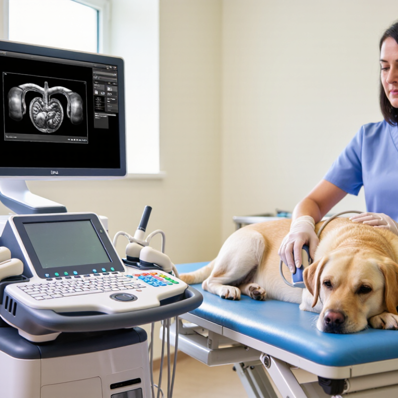
What makes a canine ultrasound machine stand out in 2026 is not its screen size. It is clinical clarity, workflow speed, and dependable handling in a busy examination room. MarketsandMarkets’ Veterinary Ultrasound Market report projects continued growth through 2028, driven by earlier diagnosis and expanding companion-animal care. That growth raises expectations. A useful system should show subtle liver textures, shallow bladder walls, and fetal movement without excessive image noise. High-frequency linear probes matter for small dogs. Curved probes remain valuable for deep abdominal views.
Real-world usability separates impressive equipment from practical equipment. A 2024 veterinary imaging survey by IMV Imaging reported that ultrasound is widely used in small-animal practice, yet training and operator confidence remain uneven. Therefore, guided presets, simple measurements, and adjustable Doppler settings can reduce avoidable errors. DICOM export, secure case storage, and fast report sharing also support better continuity between clinics and referral specialists. Battery operation helps during farm visits or crowded consultations. It should not become an excuse for weak cooling or short runtime.
AI assistance is becoming more visible, but it deserves careful skepticism. Software may outline organs or flag possible abnormalities, while a veterinarian still interprets the scan. Gartner’s 2024 AI adoption research describes rising workplace experimentation, alongside concerns about reliability and governance. That warning applies here. A machine can look advanced and still frustrate users after three examinations. In my experience, quiet fans, sealed controls, balanced probes, and a screen readable beside a nervous dog often matter more than flashy automation. Test the workflow. Record the limitations.
What Are the 2026 Top Canine Ultrasound Machines?
Key Technologies in Today’s Leading Veterinary Ultrasound Systems
The strongest canine ultrasound systems combine portability, image clarity, and dependable workflow. According to Grand View Research’s 2024 veterinary ultrasound market report, demand is rising as clinics adopt non-invasive diagnostic tools. That growth reflects a practical need: faster examinations with less patient stress.
Modern systems use harmonic imaging and spatial compound imaging to sharpen borders around the liver, kidneys, and cardiac chambers. Color and spectral Doppler help assess blood flow, while high-frequency linear probes reveal superficial structures. A 2024 report from Fortune Business Insights identifies technological advancement and portable systems as major market drivers. Still, high resolution does not guarantee a correct diagnosis. Probe pressure, patient movement, and operator skill can change the image.
Tips: Choose a system with multiple probe frequencies, quick presets, and a clear Doppler display. Test it on a restless dog, not only a calm training model. Ask whether exported images retain measurements and patient data. These details matter during referrals and follow-up examinations.
Artificial intelligence is entering veterinary imaging, but its role remains uneven. It may support measurements or flag abnormalities, yet clinical judgment remains essential. I would not select a machine because it promises automation alone. Battery performance, sealed controls, service access, and training support often matter more during a busy emergency shift. Market reports show expansion, but real-world clinic evidence is still developing. That gap deserves careful attention before purchase.
| System Profile | Typical Canine Applications | Common Transducer Configuration | Typical Frequency Range | Core Imaging Technologies | Doppler Capability | Portability and Power | Best-Fit Clinical Setting |
|---|---|---|---|---|---|---|---|
| Premium Cart-Based System | Advanced abdominal, cardiac, vascular, reproductive, and emergency examinations in small to giant-breed dogs. | Convex, micro-convex, linear, phased-array, and optional specialty probes. | Approximately 1.5–18 MHz, depending on probe type and examination depth. | High-resolution 2D imaging, tissue harmonic imaging, compound imaging, speckle-reduction processing, panoramic imaging, and elastography on selected configurations. | Color, power, spectral, and tissue Doppler; advanced cardiac measurements may be available. | Designed for a fixed examination room; mains-powered with integrated data storage and network connectivity. | Referral hospitals, specialty practices, teaching hospitals, and high-volume diagnostic centers. |
| Advanced Portable Laptop-Style System | General abdominal scanning, thoracic assessment, urinary tract examinations, reproductive workups, and focused emergency ultrasound. | Convex or micro-convex probe plus a high-frequency linear probe; phased-array cardiac probe may be supported. | Approximately 2–15 MHz. | Digital beamforming, harmonic imaging, compound imaging, adjustable focal zones, cine-loop review, and image optimization presets. | Color and spectral Doppler are commonly included; power Doppler is useful for low-flow assessment. | Usually battery-assisted and transportable; practical for room-to-room use and mobile clinical work. | General veterinary hospitals, mixed consultation rooms, mobile veterinarians, and emergency teams. |
| Compact Point-of-Care Ultrasound System | Rapid triage, abdominal fluid checks, bladder evaluation, lung sliding, pleural fluid detection, and basic cardiac screening. | One multipurpose micro-convex or phased-array probe; linear probe may be optional. | Approximately 2–12 MHz. | B-mode, M-mode, quick presets, freeze and cine-loop functions, and simplified workflow controls. | Color Doppler may be available; spectral Doppler is more limited than on premium systems. | Highly portable, rechargeable, and suitable for bedside, treatment-room, or field assessment. | Primary-care clinics, emergency care, shelter medicine, ambulatory practice, and rapid triage. |
| Tablet-Connected Probe System | Focused abdominal, lung, bladder, musculoskeletal, and procedural guidance examinations. | Wireless or cable-connected linear, convex, or phased-array probe selected for the intended examination. | Approximately 2–15 MHz, depending on probe design. | Software-based image optimization, measurement packages, annotations, cloud export, and remote image sharing. | Color Doppler is available on many configurations; advanced Doppler functions vary by probe and software. | Very portable; depends on a compatible tablet or mobile device and may require separate charging management. | Mobile practices, farm calls, home visits, outpatient rooms, and space-limited clinics. |
| Dedicated Cardiac Ultrasound System | Echocardiography, chamber measurements, valve assessment, congenital heart screening, and cardiac follow-up. | Phased-array cardiac probe, sector probe, and optional micro-convex probe for smaller dogs. | Approximately 1.5–10 MHz. | High-frame-rate 2D imaging, M-mode, harmonic imaging, optimized cardiac presets, and quantitative measurement tools. | Color, pulsed-wave, continuous-wave, and tissue Doppler are important features for comprehensive cardiac studies. | Generally room-based, with a stable cart or console preferred for extended cardiac examinations. | Veterinary cardiology services, referral centers, and practices performing frequent echocardiograms. |
| Reproductive and Breeding-Focused System | Pregnancy confirmation, fetal viability checks, reproductive tract assessment, and breeding management. | Micro-convex or convex probe, with a linear probe for superficial structures. | Approximately 3–12 MHz. | B-mode imaging, harmonic imaging, cine-loop review, obstetric measurements, and image annotation tools. | Color and spectral Doppler can assist with fetal and uterine blood-flow evaluation when clinically indicated. | Portable or compact cart formats are common for breeding facilities and farm or kennel visits. | Reproductive practices, breeding kennels, referral reproduction services, and ambulatory veterinary work. |
The top canine ultrasound machines in 2026 differ less by headline features than by clinical balance. A busy veterinary team needs clear images, quick controls, and dependable probes. High-frequency linear probes can reveal skin, tendons, and small abdominal structures. Curved probes usually provide broader views of deeper organs. A cardiac probe matters when heart motion requires careful assessment.
Image quality remains central, but it is not the only deciding factor. Strong Doppler performance helps evaluate blood flow, inflammation, and vascular changes. Sensitive color Doppler can be valuable in small vessels, although settings still require experienced adjustment. Some systems offer automated measurements or decision-support tools. These features may save time, but they cannot replace a trained interpretation.
Portability separates many leading systems. A compact unit suits farm calls, emergency rooms, and cramped examination spaces. A larger console may deliver better ergonomics during long abdominal studies. Battery duration, probe durability, and cleaning resistance deserve direct comparison. So does technical support. A cheaper machine becomes less attractive when repairs interrupt appointments. Reliability is easy to overlook.
No machine wins every case. A sharp screen can still disappoint with a poor probe. I would compare identical scans, not brochures. Test a liver image, a bladder wall, and a moving heart. Ask whether the operator can change depth, gain, and Doppler settings without hesitation. The best choice depends on patient size, examination volume, staff training, and the clinic’s actual budget.
Frequency coverage is a practical way to compare canine ultrasound systems without using company or brand names. Lower frequencies generally provide greater penetration for large dogs and abdominal examinations, while higher frequencies offer better detail for superficial structures such as tendons, joints, and small vessels. The ranges shown reflect commonly used veterinary ultrasound frequency bands.
Choosing a canine ultrasound machine starts with the patient, not the price. A compact unit suits mobile veterinarians and small clinics. It should be light enough for transport, yet strong enough for abdominal scans. A microconvex probe usually offers useful depth for most dogs. Smaller linear probes help examine tendons, superficial masses, and testicular tissue.
Large dogs Large dogs often need deeper penetration and stable image quality. In these cases, a machine with adjustable frequency ranges and reliable Doppler performance becomes valuable.
Doppler and cardiac examinations Doppler can help assess blood flow around organs, vessels, and suspected masses. For cardiac examinations, select a system with a phased-array probe and clear motion tracking.
Small dogs and puppies may require higher-frequency imaging for sharper details.
I have found that workflow matters as much as specifications. A bright screen helps during outdoor visits. Fast booting reduces stress when a restless dog is on the table. Easy image storage supports follow-up comparisons and medical records.
Training still matters. Even an advanced machine can produce misleading images with poor positioning or excessive pressure. No system is perfect.
I have also underestimated probe comfort during long scanning sessions. Clinics should test controls, service support, and probe handling before purchase. A practical trial with different dog sizes may reveal problems that a brochure hides.
The strongest canine ultrasound machines in 2026 should deliver accurate images under real clinical pressure. A high-resolution display helps identify thin intestinal walls, small vessels, and subtle abdominal changes. Multiple transducers also matter. A microconvex probe suits general abdominal scans, while a linear probe supports superficial structures and tendon work. Sensitive Doppler imaging can reveal altered blood flow, but poor settings may create misleading signals.
Safety requires more than a low-power label. Look for clear thermal and mechanical index controls, adjustable output power, and easy access to approved scanning presets. These features support the ALARA principle, which keeps exposure as low as reasonably achievable. Veterinary teams should also check probe surfaces for cracks and confirm calibration regularly. Small faults can affect image quality. They can also waste valuable time.
Clinical efficiency depends on thoughtful workflow design. Fast boot times, programmable measurements, cine-loop review, and simple patient data entry reduce repeated steps. Secure image export helps veterinarians share findings with referral specialists. Training remains essential, even with advanced automation. A machine may suggest a measurement, but a skilled operator must verify it. No system is perfect. In practice, a bright image can still hide an incorrect angle, excessive pressure, or patient movement. The best choice combines dependable engineering, transparent service documentation, and training that matches everyday canine cases.
Clinical clarity, quick controls, and dependable handling matter most. Clarity matters most. The system should show liver texture, bladder walls, and fetal movement with limited noise.
High-frequency linear probes suit small dogs, skin, tendons, and shallow structures. Curved probes provide broader views of deep abdominal organs. A cardiac probe helps assess heart motion.
Doppler helps evaluate blood flow, inflammation, and vascular changes. Sensitive color Doppler can reveal small vessels. However, poor settings may create misleading images.
Portable systems work well during farm visits, emergencies, and crowded consultations. Larger consoles may feel better during long abdominal examinations. Battery life and cooling still require careful checking.
Software may outline organs, suggest measurements, or flag possible abnormalities. It can save time during routine work. It cannot replace trained interpretation.
Guided presets, simple measurements, adjustable Doppler controls, and fast depth changes support consistent examinations. DICOM export and secure case storage improve information sharing. Staff training still matters.
Compare identical scans instead of relying on brochures. Test a liver image, a bladder wall, and a moving heart. Noise hides detail.
Check probe durability, cleaning resistance, fan noise, screen readability, and technical support. A balanced probe reduces hand fatigue. A cheaper system may cost more when repairs interrupt appointments.
No system wins everything. The best choice depends on patient size, examination volume, staff confidence, and budget. I might overvalue flashy automation, so real workflow testing deserves more weight.
In 2026, a leading Canine Ultrasound Machine is defined by image clarity, reliable performance, user-friendly operation, and the ability to support a wide range of veterinary examinations. Advanced imaging technologies can improve tissue visualization, Doppler functions can assist with blood-flow assessment, and portable designs can make diagnostic work more flexible in clinics or mobile settings. Systems that combine responsive controls, efficient workflows, and customizable settings help veterinary professionals examine patients with greater confidence.
The best choice depends on the dog’s size, medical condition, examination type, and the clinic’s daily workload. Compact systems may suit routine checkups and field use, while more advanced platforms can support detailed abdominal, cardiac, reproductive, and emergency evaluations. Important features include high-resolution probes, dependable safety controls, ergonomic design, easy data storage, simple maintenance, and clear reporting options. By comparing performance, versatility, operating comfort, and long-term practicality, veterinary teams can select equipment that improves accuracy, patient care, and clinical efficiency.

EACENIC SCIENCE & TECHNOLOGY